
IgG Subclasses and Allotypes: Structure, Biological Functions, and Immune Mechanisms
Introduction
Immunoglobulin G (IgG) is the most abundant antibody in human serum, representing approximately 10–20% of total plasma proteins. IgG belongs to the immunoglobulin family, which includes five major classes: IgM, IgD, IgG, IgA, and IgE. These antibodies are glycoproteins composed of protein (about 82–96%) and carbohydrate components (about 4–18%), and they play a central role in adaptive immune defense.
IgG antibodies are essential for pathogen recognition, immune signaling, and activation of immune effector mechanisms. Despite their structural similarity, IgG molecules are divided into four subclasses based on differences in their heavy chain structure:
- IgG1
- IgG2
- IgG3
- IgG4
Although these subclasses share more than 90% amino acid sequence similarity, they differ in several biological properties, including:
- Antigen binding efficiency
- Immune complex formation
- Complement activation
- Interaction with immune effector cells
- Circulating half-life
- Placental transport
These structural and functional differences allow IgG subclasses to adapt immune responses to different types of pathogens and antigens.
IgG Antibody Responses and Immune Regulation
The type of antigen and its route of entry into the body influence which IgG subclass is produced during an immune response. Antibody production results from B-cell activation and class switching, a process regulated by several signals such as:
- Antigen recognition by B-cell receptors
- Activation of pattern-recognition receptors (Toll-like receptors)
- Cytokines released by immune cells
- Interactions with antigen-presenting cells and T-cells
Protein Antigens
Protein antigens generally stimulate T-cell-dependent immune responses. In these cases, B-cells present antigen fragments through MHC class II molecules, leading to antibody class switching predominantly toward:
- IgG1
- IgG3
However, IgG4 and IgE responses can also occur depending on immune conditions.
Polysaccharide Antigens
Polysaccharide antigens often trigger T-cell-independent responses, resulting mainly in IgG2 production.
Repeated exposure to certain antigens may lead to secondary class switching, generating additional antibody subclasses during the immune response.
Characteristics of the Four IgG Subclasses
IgG1
IgG1 is the most abundant IgG subclass in human serum and is primarily produced in response to soluble protein antigens and membrane proteins.
- Strong complement activation
- Efficient binding to Fc receptors
- Major mediator of immune responses against viruses and protein antigens
IgG1 deficiency can lead to reduced total IgG levels, which may cause hypogammaglobulinemia and recurrent infections.
IgG2

IgG2 antibodies play a critical role in the immune response against bacterial polysaccharide antigens, particularly those present in capsulated bacteria.
Biological Functions
- Recognition of carbohydrate antigens
- Defense against encapsulated bacteria
- Contribution to immune responses against bacterial infections
IgG2 deficiency is associated with increased susceptibility to bacterial infections, especially those caused by polysaccharide-coated pathogens.
IgG3
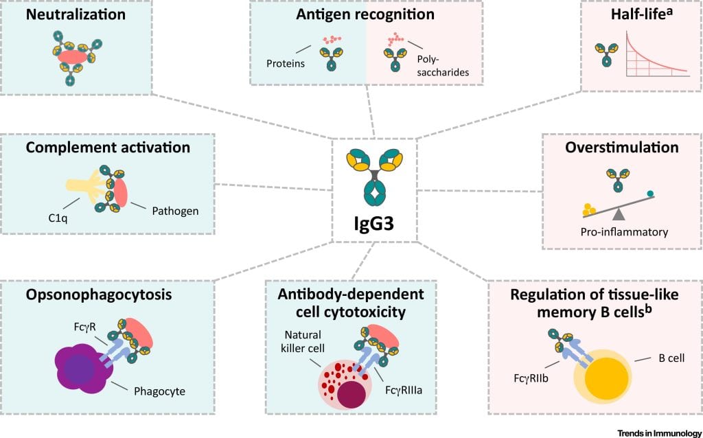
IgG3 is one of the most potent IgG subclasses for activating immune effector functions. It is particularly effective in triggering inflammatory immune responses.
Functional Properties
- Strong activation of immune effector cells
- Efficient complement activation
- Early appearance during viral infections
IgG3 antibodies often appear early in immune responses, especially during viral infections. However, this subclass generally has a shorter half-life compared with other IgG subclasses.
IgG4
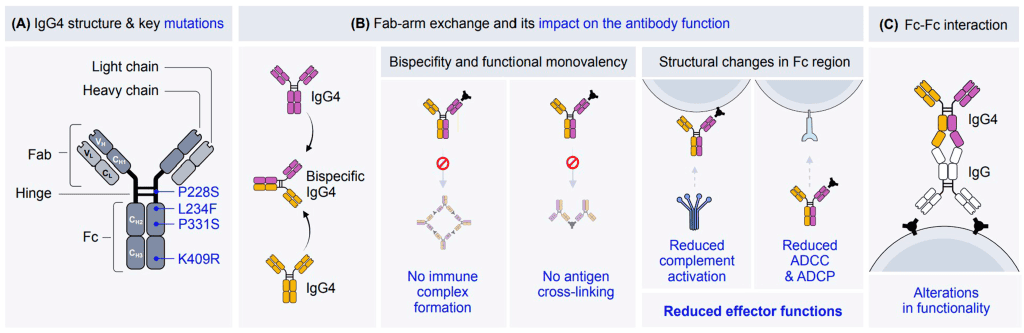
IgG4 antibodies are typically produced after repeated or long-term exposure to antigens, especially in non-infectious environments.
Common Contexts of IgG4 Production
- Chronic allergen exposure
- Long-term immunotherapy treatments
- Responses to certain therapeutic proteins
- Parasitic infections
IgG4 is often associated with immune tolerance mechanisms and may reduce excessive inflammatory responses.
Elevated IgG4 levels are also linked to IgG4-related diseases, a group of inflammatory conditions affecting multiple organs.
Structural Organization of IgG Antibodies
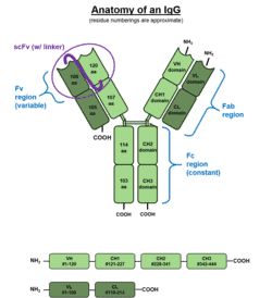
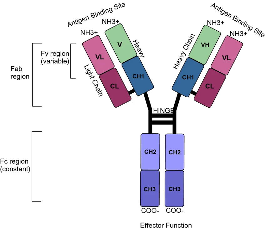
IgG molecules have a basic antibody structure composed of four polypeptide chains:
- Two identical heavy chains (γ chains)
- Two identical light chains (κ or λ)
These chains are connected by disulfide bonds, forming a characteristic Y-shaped structure.
Structural Domains
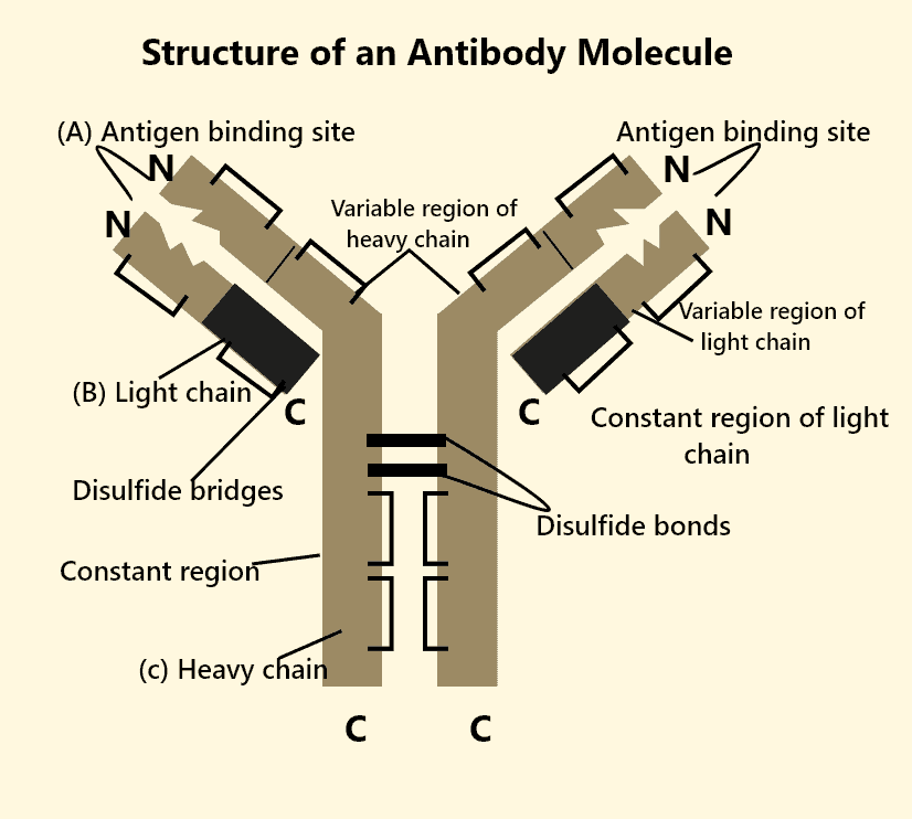
Each heavy chain contains:
- A variable domain (VH) responsible for antigen recognition
- Three constant domains (CH1, CH2, CH3) involved in immune signaling
Light chains contain:
- A variable domain (VL)
- A constant domain (CL)
Together, the variable regions of heavy and light chains form the antigen-binding site.
The Fab and Fc Regions
IgG antibodies are functionally divided into two major regions.
Fab Region (Fragment Antigen Binding)
The Fab region contains the antigen-binding site, allowing antibodies to recognize and bind specific targets such as:
- Pathogens
- Toxins
- Foreign proteins
Fc Region (Fragment Crystallizable)
The Fc region interacts with immune receptors and complement proteins, enabling antibodies to activate immune defense mechanisms.
This region is responsible for:
- Complement activation
- Binding to Fc receptors
- Immune cell activation
- Antibody-dependent cytotoxicity
Structural Variation in the Hinge Region
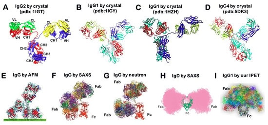
The hinge region connects the Fab arms with the Fc domain and provides structural flexibility to the antibody molecule.
The length and flexibility of the hinge region differ among IgG subclasses:
Subclass and Hinge Characteristics
IgG1 : Flexible hinge
IgG2 : Short and rigid hinge
IgG3 : Very long hinge region
IgG4 : Intermediate flexibility
- Antigen accessibility
- Immune complex formation
- Complement activation
- Interaction with immune receptors
IgG3, for example, has the longest hinge region, allowing greater flexibility but also contributing to its higher molecular weight.
IgG Allotypes and Genetic Variability
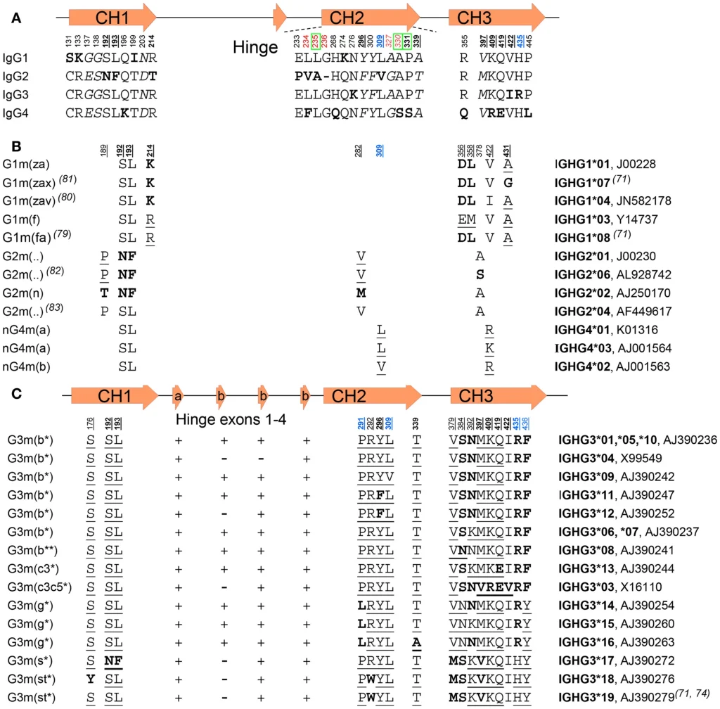
In addition to subclass differences, IgG molecules also exhibit genetic polymorphisms known as allotypes. These variations arise from allelic differences in immunoglobulin genes.
Allotypes can vary between:
- Individuals
- Populations
- Ethnic groups
These polymorphisms may influence:
- Antibody expression levels
- Binding to immune receptors
- Antibody half-life
- Immune response efficiency
Genetic variation in IgG molecules has historically been used in population genetics and forensic identification.
Glycosylation and Antibody Function
IgG molecules contain glycan structures attached to the Fc region, typically at asparagine position 297 (N297).
These carbohydrate structures influence:
- Antibody stability
- Receptor binding
- Immune activation
Different glycosylation patterns include:
- Fucosylation
- Galactosylation
- Sialylation
- Bisecting N-acetylglucosamine
Changes in glycosylation patterns can significantly affect antibody-mediated immune responses.
Effector Mechanisms of IgG Antibodies
IgG antibodies connect the adaptive immune system with the innate immune system by activating several effector mechanisms.
Major IgG-mediated immune functions include:
Complement Activation
IgG antibodies can activate the classical complement pathway, leading to:
- Opsonization of pathogens
- Membrane attack complex formation
- Pathogen destruction
IgG1 and IgG3 are the most effective subclasses in complement activation.
Fc Receptor Binding
The Fc region of IgG interacts with Fc gamma receptors (FcγR) on immune cells such as:
- Macrophages
- Neutrophils
- Natural killer cells
- Dendritic cells
These interactions trigger several immune processes including:
- Phagocytosis
- Antibody-dependent cellular cytotoxicity (ADCC)
- Cytokine release
IgG Transport and the FcRn Receptor
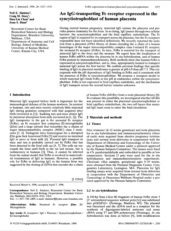
IgG antibodies have an unusually long half-life of approximately three weeks in circulation. This is regulated by the neonatal Fc receptor (FcRn).
FcRn performs several key biological functions:
- Recycling IgG antibodies in circulation
- Transporting maternal antibodies across the placenta
- Transferring IgG across mucosal barriers
This receptor plays an important role in maintaining long-term antibody immunity.
Conclusion
IgG antibodies represent a highly specialized component of the human immune system. The presence of four distinct subclasses, genetic allotypes, and structural variations enables IgG molecules to perform a wide range of immune functions.
Structural elements such as the hinge region, Fc domain, and glycosylation patterns strongly influence antibody activity, including complement activation, receptor binding, and immune regulation.
Understanding the structural and functional diversity of IgG antibodies is essential for advancing immunology research, vaccine development, and antibody-based therapies. Continuous progress in molecular and structural immunology will further improve the design of therapeutic antibodies and precision immunotherapies.